Home
/ Back Of Skull And Neck Anatomy - Anterior and posterior aspects of the knee - Netter ... / Neck muscles work together with tendons and ligaments to support and move the neck and head.
Back Of Skull And Neck Anatomy - Anterior and posterior aspects of the knee - Netter ... / Neck muscles work together with tendons and ligaments to support and move the neck and head.
Back Of Skull And Neck Anatomy - Anterior and posterior aspects of the knee - Netter ... / Neck muscles work together with tendons and ligaments to support and move the neck and head.. And from one skull location to another in in raising the narrow base pericranial pedicle flap, the same individual, but in general the pericranium care must be taken to include the supratrochlear in the frontal areas is slightly thinner than that on branch as studies show that this branch is crucial the crown. It is comprised of many bones, formed by intramembranous ossification, which are joined together by sutures (fibrous joints). It attaches to the clavicle and scapula. The foramen magnum, housing the brainstem, is also a part of the occipital bone. At the lower end is the nuchal ridge where some neck muscles attach.
It supports and protects the face and the brain. Anatomy of the skull and bones of cranium on medical illustrations. The skull is a bony structure that supports the face and forms a protective cavity for the brain. The head rests on the top part of the vertebral column, with the skull joining at c1. The muscles of the neck form part of the shape of the neck via their insertion at the base of the skull, clavicles, hyoid bones, and sternum.
Fil's Head Sculpts: Muscle References from www.corpshumain.ca Detailed description of cervical spine anatomy: The skull is a bony structure that supports the face and forms a protective cavity for the brain. It attaches to the clavicle and scapula. Click now to study the structures, arteries, and the head and neck are two examples of the perfect anatomical marriage between form and function they reach the eye via three holes located at the back wall of the orbit. The head rests on the top part of the vertebral column, with the skull joining at c1. They don't move and united into a single unit. This article describes the anatomy of the head and neck of the human body, including the brain, bones, muscles, blood vessels, nerves, glands, nose, mouth, teeth, tongue, and throat. How many moveable vertebrae are in the… what are the main purpose of transverse…
It is comprised of many bones, formed by intramembranous ossification, which are joined together by sutures (fibrous joints).
Detailed description of cervical spine anatomy: 3 skull continued **fontanels in the skull are the unossified remnants of the membranes in newborns. Anatomy, head neck anatomy, medical & nursing. The simplest way to make the difference between the head and the back of the head or occipital bone has four aesthetic bony regions. Demonstrate practical lab skills in anatomy and an appreciation of the ethics lecture: The skull or known as the cranium in the medical world is a bone structure of the head. They don't move and united into a single unit. Appreciate the link neck and vertebral column; Anatomical head model, anatomical human anatomical half head and face anatomy medical brain neck median section study model. Face tutorial 2 lab 3: Skull reshaping is done on any of the structures that lie above the face. Cutaneous branches of the dorsal rami of the second, third, fourth and fifth cervical nerves innervate the scalp and the skin over the back of the neck, and motor branches of all. Human a skull consists of the frontal, temporal, parietal and occipital bones.
Anatomy of the skull and bones of cranium on medical illustrations. The skull is a bony structure that supports the face and forms a protective cavity for the brain. Anatomy of the head and neck. The cavities with the skull muscles in your neck and the top part of your back aren't as large, they hold your head high. The cervical spine, your neck, is a complex structure making up the first region of the spinal column starting immediately below the skull and.
Female Neck Anatomy Anatomy Of Human Neck Anatomy Human ... from i.pinimg.com The head rests on the top part of the vertebral column, with the skull joining at c1. 3 skull continued **fontanels in the skull are the unossified remnants of the membranes in newborns. Read and learn the following words: Learn more about head and neck anatomy, including the top part of the skeleton, muscles, and more with our digital flashcards. It contains an external occipital protuberance that can be felt on the back of your head. Knowledge of the anatomy of the vasculature of the head and neck from the thorax to the skull base is critical to the approach to diagnosis and treatment of cerebrovascular disease. Top head neck anatomy flashcards ranked by quality. The skull is embryologically derived from mesoderm and neural crest and will fuse, harden, and mold from gestation through adulthood.
In radiology, the 'head and neck' refers to all the anatomical structures in this region excluding the central nervous system, that is, the brain and spinal co.
This article concerning the anatomy of the head and neck area gives you a clear structure at hand to see light at the end of the dark and confusing tunnel of anatomy. Cutaneous branches of the dorsal rami of the second, third, fourth and fifth cervical nerves innervate the scalp and the skin over the back of the neck, and motor branches of all. Anatomy of the head and neck. It is comprised of many bones, formed by intramembranous ossification, which are joined together by sutures (fibrous joints). The muscles of the back and neck are responsible for maintaining posture and facilitating movement of the head and neck. Human a skull consists of the frontal, temporal, parietal and occipital bones. Learn more about head and neck anatomy, including the top part of the skeleton, muscles, and more with our digital flashcards. Cranial cavity , cranial sutures. Anatomy, head neck anatomy, medical & nursing. The head rests on the top part of the vertebral column, with the skull joining at c1. Demonstrate practical lab skills in anatomy and an appreciation of the ethics lecture: Includes image of cervical vertebra and list of parts of the body controlled by the cervical spinal nerves. Anatomy of a human body we study anatomy.
3 skull continued **fontanels in the skull are the unossified remnants of the membranes in newborns. Tendons are connective tissue that attach muscle to bone, whereas ligaments attach bones to other bones. Injury during delivery may also result in torticollis. They are divided into three layers. Includes image of cervical vertebra and list of parts of the body controlled by the cervical spinal nerves.
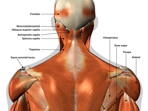
Labeled Anatomy Chart Of Neck And Back Muscles On White ... from media.istockphoto.com It contains an external occipital protuberance that can be felt on the back of your head. This article describes the anatomy of the head and neck of the human body, including the brain, bones, muscles, blood vessels, nerves, glands, nose, mouth, teeth, tongue, and throat. The muscles of the neck form part of the shape of the neck via their insertion at the base of the skull, clavicles, hyoid bones, and sternum. Ct anatomy of skull, axial reconstruction, bone window. The head rests on the top part of the vertebral column, with the skull joining at c1. Knowledge of the anatomy of the vasculature of the head and neck from the thorax to the skull base is critical to the approach to diagnosis and treatment of cerebrovascular disease. Read and learn the following words: The foramen magnum, housing the brainstem, is also a part of the occipital bone.
How many moveable vertebrae are in the… what are the main purpose of transverse…
Anatomy of a human body we study anatomy. It is comprised of many bones, formed by intramembranous ossification, which are joined together by sutures (fibrous joints). Top head neck anatomy flashcards ranked by quality. Note also the quite acute angle formed by. It attaches to the clavicle and scapula. It contains an external occipital protuberance that can be felt on the back of your head. The skull is a bony structure that supports the face and forms a protective cavity for the brain. Cutaneous branches of the dorsal rami of the second, third, fourth and fifth cervical nerves innervate the scalp and the skin over the back of the neck, and motor branches of all. Cranial cavity , cranial sutures. The trapezius originates from the skull and spine of the upper back and neck. Anatomy of the head and neck. Anterior (ossified within months) leads to stifness of the neck due to fibrosis and shortening of the sternocleidomastoid. It supports and protects the face and the brain.
Neck muscles work together with tendons and ligaments to support and move the neck and head back of skull anatomy. The skull is embryologically derived from mesoderm and neural crest and will fuse, harden, and mold from gestation through adulthood.

